
BIO340 Laboratory Guide #3
ACTIVE TRANSPORT IN
THE FROG SKIN
Frogs live in freshwater environments which are strongly hypotonic relative to their internal bodily fluids. For a typical frog, the cells, blood, and extracellular fluid are about 300 mOsm, while the external water is 40-50 mOsm. Frog skin is thin enough to permit cutaneous respiration, but one consequence of this and the hypotonicity of the aquatic environment is that water diffuses in quantity across the skin and into the frog. To compensate for this water influx, which may amount to as much as 30% of the body weight per day, frogs produce copious amounts of dilute urine. You can easily confirm this simply by grabbing a frog, or handling it roughly. There is a loss of sodium in this urine which is compensated for both by Na+ uptake from ingested food, and by the active uptake of Na+ from pond water through the epidermis. During winter months in temperate climates frogs remain submerged and feed very little, so virtually all sodium replenishment is through epithelial uptake. Epithelial cells of the frog skin thus perform some of the same functions as tubule cells of mammalian kidneys. The similarity of functions and mechanisms extends to parallel responses to drugs and hormones (e.g. mercurial diuretics, ADH, etc.).
The active transport of sodium across the plasma membrane appears to be an almost universal property of eukaryotic cells. Cell membranes contain "pumps" - ATP-dependent protein complexes which move specific ions "uphill" across the membrane, against their normal concentration gradients. The sodium/potassium pumps move sodium out of the cell and potassium into the cell, at significant metabolic expense. As a result, sodium becomes the dominant cation of the extracellular fluid (ECF), while potassium becomes the dominant cation of the intracellular fluid.
At first glance, this exchange of cations would not seem to generate any net current (movement of charges across the membrane), or any membrane potential (electromotive force resulting from unequal distribution of charge across the membrane). However, both sodium and potassium ions tend to leak back across the membrane down their concentration gradients. Since cell membranes are generally much more permeable to K+ than to Na+, K+ has a stronger tendency to leak back out of the cell. This process indirectly generates the so called Nernstian component of the "resting" membrane potential. Furthermore, most Na/K pumps do not exchange ions 1:1. Vertebrate pumps generally pump 3 Na+ ions out for every 2 K+ ions pumped into the cell. This makes the vertebrate Na/K pump electrogenic, i.e. it directly produces an unequal distribution of charge and hence a small additional transmembrane potential, which also contributes to the resting membrane potential.
The frog epidermis is a multilayer stratified epithelial tissue (like the human epidermis). However, it can be treated as if it were a single cell thick for discussions of epithelial transport. As described in your text, Na/K pumps are concentrated on the basal (serosal, internal) side of the frog skin, in fact on the basal side of the basal cell layer. At this surface, sodium is pumped from the epithelium into the ECF of the frog. This lowers the intracellular sodium concentration of the epithelial cells to the point where sodium flows down its gradient from the external pond water, through the relatively sodium permeable apical membranes and into the epithelial cells. Potassium is pumped into the cells by the Na/K pumps at the basal surfaces, but its relatively high permeance allows it to flow back down its concentration gradient into both the pond water and the ECF. If sodium were the only ion with a net flux across the membrane, an electric charge (positive inside) would rapidly build up across the epithelium, forming an effective electrical gradient which would block further sodium transport. However, in the frog skin, chloride ions flow "passively" across the epithelium, from outside to inside, generating a natural countercurrent (they flow down the electrical gradient established by the Na/K pump). The primary route for this flow of Cl- is paracellular, i.e. through the spaces between the cells. This chloride flux results in a current which exactly matches in amplitude the current carried by cations. In other words, although chloride and sodium fluxes are both into the frog, there is no net current flow, because the net cationic and anionic currents flow in opposite directions (because cations and anions have opposite charges). Consequently a constant, steady-state electrical potential is maintained across metabolically active frog skin. The amplitude of this potential, as well as the amplitudes of the cationic and anionic currents are determined by the ionic makeup of the solution of either side of the frog skin, and the activity level of the sodium/potassium pumps.
Because the frog skin can be easily isolated and studied, it has attracted the attention of physiologists for many years, and has become a standard preparation for introductory physiology labs. We will be using a simple and classical apparatus, the Ussing chamber, to study sodium active transport. This apparatus consists basically of a isolated piece of frog skin separating two fluid compartments. Sodium active transport generates a potential difference across the frog skin which can be measured by immersing a suitable electrode (in our case a Ag/AgCl post) in each compartment. We will look first at this direct electrophysiological effect of epithelial sodium transport. We will then be applying an electrical countercurrent to "short-circuit" this potential and, in the process, isolate and measure the actual current generated by the all of the sodium pumps in a unit of membrane area. We will then study the ionic specificity and pharmacological dependence of epithelial transport. Finally, we will look at the metabolic requirements of active transport.
The Ussing chamber compartment volumes are large, relative to the surface area of the frog skin and the rate of pumping. This means that the currents produced by active transport are not expected to appreciably affect the concentrations in either compartment, over the time course of this experiment. This is very different from the situation in the interior of a cell, where the Na/K pump establishes and maintains and internal ionic milieu radically different from that of the extracellular fluid.
It will be helpful for you to keep in mind throughout this course that an immeasurably small flux of ions (moles/sec) can carry a large and easily measured current (coulombs/sec = amps). The relationship between flux and current is the same as that between concentration and charge, and is expressed by the Faraday constant (F = 96,500 coulombs/mole for a monovalent cation). Thus, a current measured in microamps (1A = 10-6 amps) corresponds to a net transport measured in tens of picomoles (10-12 moles) per second. A 1 A current carried by a monovalent ion would therefore require ~105 seconds (~28 hours) to change the concentration of a 1 ml volume by 1 mM.
Additional Reading:
Pages 90-98,282-283 in Animal Physiology (2nd Ed.) by Hill, Wyse, and Anderson.Box 4.1 on freshwater fish gills works for frog skin as well.
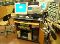
I. EQUIPMENT SETUP
For this lab, we will be using the PowerLab Digital Voltmeter (DVM - accessible through Chart) to monitor the potential between the two compartments of the Ussing chamber. In the "short-circuit" configuration we will add a second "circuit" to the Ussing chamber to provide a countercurrent flow. This circuit will consist of a DC source (2 C batteries), a set of potentiometers to regulate the electrical current flow, and a microammeter to measure this imposed current flow.
A small digression on electrodes is in order here. Assuming we have two wire leads coming off of a recording or stimulating circuit, what is an appropriate way to make contact with the fluid on the two sides of the membrane? On first thought, it might seem sufficient to just immerse the free end of each wire in the fluid. If this is done, a potential difference will be observed between the wires, but it will be erratic and bear little relationship to the actual potential difference between the two compartments. When a bare metal electrode is immersed in a saline solution, an electrical potential is established between the metal and the solution, due to the metal yielding ions to the solution, or the solution plating ions onto the metal. This is called electrode or junction potential, constitutes a variable battery, and is the bane of electrophysiology. If the junction potentials at the two electrodes were constant and identical, they would cancel out in the overall potential measurement. In order for this to happen, both the conditions at the surface of each electrode, and the ionic makeup of the solution surrounding the electrode would have to be constant and identical. However, as current flows in a single direction through each junction, the properties of the junction change, e.g. as a result of electroplating of ions onto the electrode. This process is compounded when electrodes are used to pass externally applied direct currents. Under such conditions, bare metal electrodes can rapidly "polarize" and block further current flow. To minimize these effects, we will be using metallic silver electrodes which have been electrically coated with a layer of silver chloride. When placed into a NaCl solution, for example, the Ag/AgCl/NaCl interface remains fairly stable and can accurately record and pass direct (unchanging) currents.
The equipment needed for this experiment includes an Ussing chamber, Ag/AgCl electrodes, a microammeter, a potentiometer box, two dry cell batteries, and a PowerLab/PC recording station.
Equipment Setup Procedure:
1) Rinse out the two compartments of the Ussing chamber with tap water, followed by distilled water, followed by a small amount of frog Ringer's. Avoid touching the inner surface of the Ussing chamber blocks. Clean any crusted salt off of the base clamp and the spacer blocks.
2) Obtain four chlorided silver post electrodes from the instructor. Keep them immersed in the frog Ringer's solution in a small beaker until you are ready to record.
3) Launch Chart. Turn off all channels except Channel 1. Set Channel 1 for bipolar recording (activate both the positive and negative input leads) at a sampling rate of about 4 per second and with an amplifier display range of 200mV.
4) Select DVM:Channel 1 from the Windows menu. Enlarge the DVM window by pulling down on the lower right corner. The DVM will simply reflect the voltage present across the + Channel 1 input leads whenever Chart is recording.
5) Assemble the Ussing chamber without any membrane. Fill the chamber with 9.0cc of frog Ringer's. Place a Ag/AgCl electrode in the innermost holes on each side of the chamber. Connect these electrodes to the "+" and "-" inputs of CH1 on the PowerLab, connecting "+" to side A and "-" to side B of the chamber.
6) Record the DVM electrical potential in mV between the two electrodes. This is a potential is produced by differences between the two electrodes and will have to be subtracted from all subsequent voltage measurements to isolate potentials generated by the frog skin itself. This "electrode potential" is also the "zero" or baseline voltage at which you will be holding the preparation for Section III parts B-E. You may want to try various pairs of your four electrodes and select the pair which produces the smallest baseline electrode voltage.
7) Label the dry ends of your two electrodes A and B with small pieces of tape, unclip them, then return them to their original Ringer's container. If you fail to unclip them at this point, the "mystery metal" of the clips will rapidly electroplate onto the electrodes and ruin them. Drain and rinse the Ussing chamber.
II. DISSECTION AND MOUNTING OF THE FROG SKIN
For this and future experiments involving animal surgery, always wear surgical gloves, change your gloves whenever they become torn or contaminated. As soon as you are done handing the frog skin, remove your gloves and wash your hands thoroughly .
Procedure:
1) Obtain a decapitated or pithed frog from the instructor.
2) Use your scissors to remove a piece of skin from the ventral surface of the frog at least 2.5cm in diameter.
3) Return the remains of your frog to an ice chest when you are through, or give it to another group of students.
4) Carefully mount the frog skin between the blocks off the Ussing chamber, using just enough clamp pressure to hold it in place. Make sure that there are no holes or gaps in the skin which could allow direct flow of fluids between the two compartments. The skin should be mounted so that the exterior (mucosal, apical, green) surface faces the B compartment and the interior (serosal, basal, white) surface faces the A compartment. If you get the polarity reversed, do NOT remount the frog skin, just relabel the compartments.
5) Fill one chamber with 4.5cc of frog Ringer's and check for leaks. Then fill the other chamber with 4.5cc of frog Ringer's. Slowly bubble 10cc of air into each compartment of the Ussing chamber, using the 10cc syringe and the holes farthest from the epithelium.
III. RECORDING FROM THE FROG SKIN
The voltage recorded across the frog skin (transepithelial potential) will reflect not only the active transport of cations (predominantly Na+), but also the passive flow of anions (predominantly Cl-) across the epithelium. Since Cl- ion concentrations are equal on the two sides of the membrane, and assuming that Cl- ions are not actively pumped, any Cl- fluxes must be driven by the electrical potential across the membrane. If we could eliminate this transepithelial potential, by applying an electrical countercurrent, Cl- ions would cease to flow. Furthermore, the electrical countercurrent which we apply across the membrane to hold it at a zero potential, would exactly match (with opposite polarity) the net current due to active transport of cations. This follows from Kirchoff's Current Laws - essentially stating that the same current must flow at all points in a simple, unbranched circuit.
In order to "short-circuit" the epithelium, pin it to zero potential, and measure the current required to do so we must construct a second circuit across the epithelium, containing a steady voltage source, a potentiometer (variable resistor) to regulate the current in this circuit, and a microammeter to measure the current. According to Ohm's law, for resistive current:
V = IR
where V is the potential in volts, I is the current in amps, and R is the resistance in ohms. With a fixed voltage source V (in our case a battery), the current I flowing through the circuit can be varied by changing the resistance R at the potentiometer. We will use this short-circuit countercurrent to quantify sodium transport for the bulk of this laboratory exercise.
As you work through the following procedures, keep three things in mind:
1) Fluid levels in the two chambers must be exactly equal, so that there is no hydrostatic pressure between the two chambers whenever you are recording an electrical potential.
2) At least once every 5 minutes during the following experiments you must slowly bubble 10cc of air into each compartment of the Ussing chamber, to keep the epithelium oxygenated and allow active transport processes to continue.
3) This preparation has a limited viability. Work as rapidly as you can. Answer short questions as you go, and make sure that you have sufficient data for the required calculations. However, wait to perform the longer calculations until after the experiment is complete.
A. Measuring the Electrical Potential Generated by Active Transport
1) Return your two recording electrodes to the chamber and connect them to the PowerLab box, as before. Be careful that the electrodes never touch the frog skin.
2) Activate the DVM, by clicking on the Start button.
Q1: Which side of the skin is positive relative to the other? What does this tell you about the likely direction of sodium pumping when there are no ionic gradients between the two chambers.
3) Inactivate the DVM when you are through.
4) Remember to periodically bubble air through the two compartments.
B. Measuring the Active Transport Current
1) Insert the second pair of electrodes in the central (smallest bore) holes of each compartment of the Ussing chamber. Make sure that these electrodes do not contact the other electrode pair. Using the wires with alligator clips assemble a circuit running from one of these electrodes, through the battery, through the potentiometer box, through the microammeter, and to the other electrode.
2) Adjust the potentiometers to produce the lowest possible current flow, as measured by the microammeter.
3) Again, remember to periodically bubble air through the two compartments.
4) Now activate the DVM. Adjust the potentiometers to increase the countercurrent until the voltage across the frog skin is zero. Remember that you must subtract the DC electrode voltage from the DVM reading to obtain the true voltage across the skin. If increasing the applied current increases the potential, reverse the connections to your two current-supplying electrodes.
5) Record the current required to produce a zero transepithelial potential (again, remember to take into account your measured baseline electrode potential).
6) Inactivate the DVM when you are through, and drop the applied countercurrent to its lowest value.
Q2: Does the shortcircuit countercurrent flow in the same or opposite direction as the active transport current? Explain why.
Assume that this short circuit current is required to supply one electron for every net monovalent cation pumped across the membrane. Assume further that Na+ is the only cation effectively displaced by the pump. The net transport rate of Na+ across the epithelium may be calculated from the following definitions:
1 microampere = 10-6 amperes
1 ampere = 1 coulomb/second
96,500 coulombs = 1 mole of monovalent charge carriers
(either electrons, Na+ ions, or K+ ions)
It follows that each �amp of short circuit current is equivalent to 1.04 x 10⁻¹¹ moles per second of net cation transport across the membrane. Do the calculations for the following questions after you have finished with the experiment:
Q3: Given your recorded short-circuit current, calculate the net Na+flux (in moles/sec) across the epithelium in your Ussing chamber.
Q4: The diameter of the main Ussing chamber is 12.5mm. Calculate the net Na+ flux per unit surface area (moles/sec mm2) due to active transport.
Q5: Given a volume of 4.5ml in each compartment of the Ussing chamber, how long would it take to change the concentration of Na+ on either side of the membrane by 1mM? Does your result justify our initial assumption that ionic concentrations in the two chambers could be treated as invariant?
C. Pumping Against Ionic Gradients and Cation Specificity of the Pump
In the experiments so far, the concentration of ions on each side of the frog skin has been identical. However, in the living frog, sodium is pumped against a substantial gradient. We will verify that sodium is carrying the active transport current and study the concentration dependence by substituting K+ for Na+ on the external side of the epithelium, while keeping the transepithelial potential short-circuited to zero.
1) Activate the DVM and record the short-circuit current necessary to zero the transepithelial potential.
2) Now, using 3cc syringes, withdraw 2.25 cc of Ringer's from the outside compartment of the Ussing chamber and immediately replace it with 2.25cc of K-substituted Ringer's (a frog Ringer's solution in which NaCl has been replaced with KCl). Bubble air through the compartment to oxygenate and mix the solution. This solution will now contain 1/2 of the initial Na+ concentration.
3) Record the short circuit current under this condition.
4) Repeat steps 2&3 to produce outside Na+ concentrations of 1/4, 1/8, 1/16, and 1/32 of the original concentration, recording the required short-circuit current under each condition.
5) When you have made the final measurement, inactivate the DVM and set the countercurrent to the lowest value. Remove both pairs of electrodes and gently flush both compartments of the chamber out with standard frog Ringer's. Do not tip the chamber over, simply extract the fluid in each side with a syringe and gently replace it with fresh frog Ringer's. Repeat the flushing process at least twice.
6) Immediately refill both sides with 4.5ml of standard frog Ringer's and return the electrodes to the chamber.
Q6: Is the pump ion-selective, i.e. are sodium and potassium equivalent vis-a-vis the pump?
Q7: Can Na+ be transported against a concentration gradient?
Q8: Does your graph provide any indication that passive Na+ diffusion plays a significant role, i.e. is there a significant Na+ "leak" across the epithelium?
D. Poisoning the Pump
Since ancient times, many cultures have used dried and powdered leaves from Digitalis (foxglove) plants to stimulate the heart and increase cardiac output. The active ingredients in these leaves are the cardiotonic glycosides digitoxin and digoxin, which depolarize cardiac cells by partially inactivating Na/K pumps. We will be using ouabain, a related, naturally occurring compound to partially block active transport. Ouabain is produced by the adrenal cortex of many vertebrates, but is commercially isolated from the seeds of the plant Strophanthus grastrus.
OUABAIN IS A POISON. WE WILL BE USING EXTREMELY SMALL AMOUNTS AT LOW CONCENTRATIONS WHICH SHOULD POSE NO REAL DANGER TO YOU. HOWEVER, HANDLE IT CAREFULLY. IF YOU SPILL IT ON THE TABLE OR YOUR SELF, CLEAN IT UP/OFF IMMEDIATELY AND NOTIFY THE INSTRUCTOR. DISPOSE OF USED PIPETTE TIPS IN THE SPECIALLY MARKED HAZARDOUS WASTE CONTAINER.
1) Activate the DVM and record the short-circuit current necessary to zero the transepithelial potential.
2) Using a pipetman, add 10 �l of ouabain solution only to the outside compartment of the chamber and 10 �l of frog Ringer's to the inside compartment to balance the volume.
3) Record the short-circuit current at one minute intervals for the next 20 minutes. After the final measurement inactivate the DVM
.
4) Using the syringe labeled "Ouabain" carefully empty each side of the chamber and transfer the contents to the container marked "Hazardous: Ouabain Waste". Refill the chambers with standard frog Ringer's. Repeat this process twice more, transferring the waste to the ouabain waste container each time.
5) Remeasure the short circuit current to confirm that the ouabain effect has been completely washed out. If the original rate of sodium transport has not been restored, wait five minutes, then test again. If the original rate of sodium transport has still not been restored, repeat the flushing process.
6) Repeat steps 2-5. but this time add the 10 �l of ouabain solution only to the inside compartment and 10 �l of frog Ringer's to the outside compartment. Flush and refill the chamber with standard frog Ringer's when you are through, transferring all rinsate to the Ouabain waste container.
7) Inactivate the DVM when you are through.
Q9: Do your ouabain results confirm the idea that the active transport across the epithelium results from Na/K pumping?
Q10: Do your ouabain results provide any information about the location in the epithelium of the Na/K pumps?
E. Metabolic Requirements of the Pump
Active transport requires energy and imposes a metabolic load on the cell. Specifically, the Na/K pump is an ATPase, and will deplete cellular ATP reserves if they are not replenished. We will be using 2,4-dinitrophenol (DNP), a metabolic poison. DNP is a hydrogen ionophore which inserts itself into the mitochondrial inner membrane, allows the transmembrane H+ gradient to dissipate, and therefore uncouples the oxidative phosphorylation process of cellular respiration, which phosphorylates ADP to ATP.
DNP IS A CELLULAR METABOLIC POISON AND IS CLASSIFIED BY THE ENVIRONMENTAL PROTECTION AGENCY AS AN EXTREMELY HAZARDOUS SUBSTANCE. IT MUST BE HANDLED CAREFULLY AND EXACTLY AS DESCRIBED BELOW. IF YOU SPILL IT ON THE TABLE OR YOUR SELF, CLEAN IT UP/OFF IMMEDIATELY AND NOTIFY THE INSTRUCTOR. DISPOSE OF USED PIPETTE TIPS IN THE SPECIALLY MARKED HAZARDOUS WASTE CONTAINER.
1) Activate the DVM and record the short-circuit current necessary to zero the transepithelial potential.
2) Using a pipetman, add 10 �l of DNP solution to each compartment of the chamber.
3) Record the short-circuit current at one minute intervals for the next 20 minutes. After the final measurement inactivate the DVM.
4) Using the syringe labeled "DNP" carefully empty each side of the chamber and transfer the contents to the container marked "Hazardous: DNP Waste". Refill the chambers with standard frog Ringer's. Repeat this process twice more, transferring the waste to the ouabain waste container each time.
5) Remeasure the short circuit current to confirm whether the the DNP effect has been completely washed out. If the original rate of sodium transport has not been restored, wait five minutes, then test again. If the original rate of sodium transport has still not been restored, repeat the flushing process.
6) Inactivate the DVM when you are through
Q11: What do your results tell you about the metabolic requirements of active transport across the frog epithelium?
Q12: Could the DNP effect be washed out?
IV. PREPARATION OF THE LAB DATA SHEET
Your data sheet should include all THREE of the items described in the boxes above. Make sure that the axes of all of the graphs and print-outs are labeled and calibrated. You should certainly discuss your results and the answers to the questions with your partners and others in the lab. However, please work independently when you prepare your data sheet.