
BIO340 Laboratory Guide #2
PERMEABILITY OF THE
RED BLOOD CELL MEMBRANE
The phospholipid bilayer is the major structural element of all cell membranes. The chemical nature of this bilayer enables the membrane to form a semipermeable barrier to the extracellular environment, thereby regulating the flow of dissolved molecules and ions into and out of the cell. Some molecules can diffuse readily through the bilayer; in their case the membrane provides little real barrier at all. At the other extreme are molecules that are completely impermeant to the lipid bilayer. If these molecules are to move across the membrane, they must pass through specific channels or be carried by specific transport proteins in the membrane. The movement of water across the membrane is as important as the movement of solutes. When the concentrations of uncharged solutes on either side of the membrane are unequal, permeant solutes will diffuse across the membrane, tending to redistribute themselves such that the osmolarity (concentration of dissolved solutes) of each solute on both sides of the membrane is equal. Impermeant ions will not cross the membrane and will remain at their initial concentrations. In either case, water will diffuse across the membrane in the direction of greater total osmolarity (higher concentration of all solutes combined = lower water concentration). In simple terms, water will "follow" movement of permeant solutes or standing concentration gradients of imperment solutes into or out of cells. The flow of water into or out of the cell will lead to a change in cell volume. Because of this, cell volume changes can be used as indicators of the permeability of the cell membrane to particular solutes or the relative concentrations of imperment solutes across the membrane.
Erythrocytes, commonly called red blood cells (RBCs), constitute 30-40% of the animal connective tissue known as blood. Since mammalian RBCs are easy to obtain in large quantities, have a minimum of internal organelles, and are uniform in size and shape, they are often used to quantitatively study the generic permeability properties of animal cell membranes. The RBCs you will use in this laboratory will be taken from horse blood.
Additional Reading:
Chapter 4 in Animal Physiology (2nd Ed.) by Hill, Wyse, and Anderson.
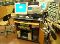
I. EQUIPMENT SETUP
Equipment needed for this lab includes a centrifuge with 15ml capped tubes, a compound microscope with slides and coverslips, a spectrophotometer with capped cuvettes, and a PC with installed Spectrometry software. The only preliminary equipment setup required for students is to locate the equipment.
II. PREPARATION OF THE RBC STOCK SOLUTION
To prepare your RBC stock solution your instructor will the blood plasma with isotonic (300mM) sucrose, an impermeant solute. To do this he will repeatedly centrifuge the blood solution to pellet the RBCs, remove the supernatant, replace the supernatant with 300mM sucrose, and resuspend the RBCs. The idea is to produce a stock solution with a hematocrit of about 35-50%, i.e. in which RBCs make up about one third to one half of the volume.
Procedure (instructor should do this no more than four hours before the start of lab - but students should read through this to be sure that they understand the procedure, ratuionale, and results):
1) Put on a pair of surgical gloves and keep them on for the entire lab. Feel free to change gloves as they become contaminated (always treat discarded gloves for this lab exercise as orange bag waste for autoclaving). Always wash hands with antibacterial soap between glove changes. Handle the blood solutions with extreme care. Immediately clean up any spills with Clorox Cleanup or a bleach solution. All blood contaminated waste goes in the autoclave bag waste container at the front of th e room.
2) Fill several 15ml centrifuge tube with defibrinated/heparinized blood, trying to avoid contaminating the outside of the tube.
3) Spin the tubes in the IEC|Centra CL2 centrifuge at 2400 rpm for 4 minutes. MAKE SURE THAT EACH CENTRIFUGE TUBE IS BALANCED WITH ANOTHER TUBE CONTAINING A IDENTICAL AMOUNT OF BLOOD, PLACED IN THE OPPOSITE SLOT.
4) Carefully remove the supernatant in each tube with a plastic pipette. Try not to disturb the RBC pellet. Then refill each tube to 1.2 ml with 300mM sucrose. This procedure will ensure that enough sucrose can be added to the tube to wash the RBCs thoroughly.
5) Cap the tube and gently re-suspend the RBCs by upending the tube several times. Avoid making bubbles in the suspension, which could damage the RBCs.
6) Repeat steps 3, 4, and 5 two more times to thoroughly wash the RBCs.
7) Spin the tubes one more time. Add just enough 300mM sucrose to bring the volume up to ~1.5 times that of the RBC pellet.
8) Combine all of the RBC suspensions and aliquot at least 5 ml to each of four student groups, with the remainder held in reserve.
This final suspension is the RBC stock suspension. Keep all tubes stored on ice. The RBCs will settle with time, so each time that you remove an aliquot from the stock suspension, make sure that it is well mixed.
III. THE ERYTHROCYTE AS AN OSMOMETER
The RBC membrane is semipermeable, meaning that it is freely permeable to some possible constituents of the surrounding solution (e.g. water), but is practically impermeable to others (e.g. sucrose). For all practical purposes, the membrane is also impermeable to all of the solutes ordinarily found inside the cell. Given these properties, the cells should show volume changes in sucrose solutions of varying osmotic concentrations, corresponding to the tonicity of those solutions. We are going to quantitatively test this hypothesis. Specifically, we would like to know if the amount of water exchange taking place between the cells and their surrounding solution can be quantitatively accounted for, in terms of established osmotic laws.
It is a common practice among both biology students and professional biologists, when confronted with complex equations, such as the ones below, to simply skim over them and hope for the best. In a physiology class, the equations are at the heart of the subject. Please go over both the math and the arguments below until you understand both the equations and their rationales.
Under any condition, the total solute concentration (C) inside the cells is equal to the total number of moles of solute (N) divided by the volume in which they are dissolved. However, this volume is not simply the total cell volume, because all cells contain materials (cytoplasmic structural elements, organelles, lipids, etc.) that take up space, but are not in solution, as well as some "osmotically inactive" water tied up by intracellular proteins. The total space occupied by these materials is termed the "osmotically inactive volume", denoted as b. If V is the total cell volume, then V - b is the cell volume in which osmotically active solutes are dissolved, and C is given by equation (1):
C = N / (V-b) or, after rearranging, N = C (V - b ) (1)
If the solutes being studied are impermeant, then there should be no movement of solutes into or out of the cells when the cells are placed in solutions of different concentrations; only water should move across the membrane. Therefore, N0 (the number of moles of solute when the cells are in isotonic saline or 300mM sucrose) should equal N when the cells are in any other impermeant solution:
N0 = N (2)
Substituting equation (1) into equation (2) gives:
C0 (V0 - b) = C (V - b) (3)
After rearranging (do this at home until you get it right), this expression becomes:
V / V0 = (1 / C) [C0 - (bC0 / V0 )] + b / V0 (4)
Equation (4) is the rather messy-looking equation for a perfect osmometer. In any experiment C0 , V0 , and b are constant, and therefore, equation (4) is also the equation for a straight line. Therefore a plot of (V / V0) vs. (1 / C) should give a straight line, with a slope equal to [C0 - (bC0 / V0 )] and a y intercept of (b / V0) , if the hypothesis is correct.
To test the hypothesis, you will equilibrate the cells with external solutions of known osmotic concentration and then measure the resultant cell volumes.
Experimental Procedure:
Work over an absorbent pad for this entire procedure. Try to avoid contaminating any other surfaces with blood. Dispose of any contaminated pads, Kinwipes, etc in the orange bag waste container. Dispose of contaminated glass capillary tubes in a red sharps container.
Wear gloves for this entire procedure. As gloves become contaminated, dispose of them in the orange bag waste container and don new gloves. Always wash your hands thoroughly with soap between glove changes.
1) Mix the RBC stock solution and carefully transfer ~1 ml from the centrifuge tube to each of two (2) 1.5ml Eppendorf tubes.
2) Using a 1000 uL pipetter, transfer 0.25 ml (250 uL)of each of the following sucrose solutions into a separate 1.5 ml Eppendorf tube, seal the cap, and label each tube with a Sharpie pen.
Sucrose Concentrations (mM): 100 200 300 600 800 1200 (tubes 1-6, respectively)
3) Similarly, add 0.25 ml (250uL) of the RBC stock suspension into each Eppendorf tube. Remember to thoroughly mix the stock suspension before you withdraw each sample. Be careful not to contaminate your stock suspension with the sucrose solutions.
4) Recap each tube and agitate for 2-3 seconds to mix the contents. Allow the cells in all of these Eppendorf tubes to to equilibrate (on ice) with the new solutions for at least 20 minutes. You can proceed to set up other sections of this lab while you are waiting.
5) Agitate each of the seven (RBC stock + six tests) tube for 2-3 seconds, then fill a sample from the tube to a clay-sealed capillary tube, holding the capillary tube horizontal. Allow enough blood to enter each capillary tube to fill about half of the tube. As you fill each capillary tube, wipe off the outside, then seal the opposite (clean) end with a clay plug. Keep carefull track of which tube is which, i.e. what the test sucrose concentration is in each tube.
6) Load your capillary tubes into the HemataStat centrifuge. The centrifuge must be balanced with two, three, or six capillary tubes before you run it. You can fill in empty slots with unused capillary tubes.
6) Centrifuge each set of the tubes through a standard 60 second run. Following the instructions on the HemataStat screen, use the slider to measure and calculate the hematocrit (Hct) as a decimal value between 0 and 1. Record each hematocrit value to your laboratory notes.
Q1: Note the color of the supernatant in each tube and be able to explain any differences that you see. One of your tubes may not have a clear supernatant. Why not? Can you still use Hct data from this tube? Why or why not?
Calculations:
Complete these calculations at the end of the laboratory period.
Produce Table 1 on an Excel spreadsheet. Your rows will correspond to the 6 test concentrations of sucrose that you mixed with your stock solutions. Your columns (A-H) will be the eight variables and constants listed below:
A) tube number (1-6)
B) Vt = volume of the test solution in ml (= 0.25)
C) Ct = osmotic concentration of the test solution (100, 200, 300, 600, 800, 1200)
D) Hcts = Hct of the RBC stock suspension (as measured for stock tube)
E) Vs = volume of the RBC stock suspension in ml (= 0.25)
F) Cs = osmotic concentration of the RBC stock suspension (= 300)
G) Hctf = Hct of the final solution (as measured for each tube 1-6)
H) Vf = volume of the final solution in ml (= 0.5)
Add two additional columns (I-K) for three sets of values which you will compute: C, 1/C, and V / V0.
For each row the calculate the theoretical value C = Cf and enter it column I, using the following equation:
Cf = {Vt Ct + [(1 - Hcts) Vs Cs]} / [(1 - Hctf) Vf] (5)
Now calculate and enter 1/C (=1/Cf) for each row and enter these values in column J.
Finally, calculate V / V0 for each row and enter these values in column K, based on the following logic. If you measured carefully and made sure that the RBC stock suspension was well mixed before withdrawing the samples, there should be an equal number of cells in each tube (unless the cells in the tube ruptured). Therefore, the hematocrits should be directly proportional to the average cell volumes. Since the concentration of isotonic sucrose (C0 ) is 300 mM, the ratio of the Hct in any tube to the Hct in 300 mM sucrose should equal the ratio V / V0. Therefore V / V0 for each sucrose solution is equal to Hcts/ Hct300.
Draw a graph of V/V0 (column K) as a function of 1/C (column J). Do not plot a point for the stock suspension. Be sure that both axes are correctly labeled and calibrated. Fit a straight line to your data points and extrapolate to infinite concentration (1/C = 0) on your graph to obtain b/V0 (the Y-intercept of your line) This is the fraction of cell volume that is osmotically inactive. Write this value and label on your graph with an arrow pointing to the point used to determine it.
Q2: Do the results of this experiment support the hypothesis that we set out to test? Explain.
IV. MICROSCOPIC APPEARANCE OF RED BLOOD CELLS
In isotonic solutions, red blood cells are biconcave disks with a remarkably uniform diameter (7mm for human RBCs). This shape allows them to fold up slightly as they squeeze in single file through capillaries. The cell membrane has a relatively fixed surface area. As the cell volume decreases in a hypertonic solution of an impermeant solute, the cell membrane wrinkles, and the RBC takes on a crenelated appearance. Conversely, as the cell volume increases in a hypotonic solution, the RBC will first swell and lose its biconcave shape, then eventually burst.
Work over an absorbent pad for this entire procedure. Try to avoid contaminating any other surfaces with blood. Dispose of any contaminated pads, Kinwipes, etc in the orange bag waste container. Dispose of contaminated glass capillary tubes in a red sharps container.
Wear gloves for this entire procedure. As gloves become contaminated, dispose of them in the orange bag waste container and don new gloves. Always wash your hands thoroughly with soap between glove changes.
In this experiment you will observe microscopically the effect of solutions of various osmotic strength on the gross appearance of RBCs. These qualitative observations of RBC behavior should lend further support to the hypothesis that the RBC has a semipermeable plasma membrane.
This is a good procedure to do while you are waiting for your cells to equilibrate in Step 4 of Section III.
Experimental Procedure:
Repeat the following procedure with 3 different concentrations of sucrose: first 300 mM, then 600 mM, then 100 mM.
1) Place a small drop of RBC stock suspension on a microscope slide.
2) Add 1 medium drop of sucrose solution directly on top of the RBC suspension.
3) Add a coverslip and examine the slide immediately through the compound microscope.
If the RBCs are crowded too closely together to see individual cells clearly, repeat the procedure with a smaller volume of RBC suspension.
Q3: Describe the appearance of the RBCs under each condition. With your knowledge of osmotic principles, discuss what has happened in each case.
V. HEMOLYSIS AS AN INDICATOR OF RELATIVE PERMEABILITY
So far you have been dealing only with RBCs suspended in solutions of an impermeant solute, namely sucrose, and subsequent movement of water across the membranes by osmosis. However, the environment of living cells may also contains a variety of permeant solutes which can pass through the membrane, and it is time to deal with them. For the sake of simplicity we will consider only solutions in which the solute concentration equals the total concentration of soluble substances inside the cell (300mOsm for cells equilibrated to 300mM sucrose). Note that while a 300 mM solution of a permeant ion is, by definition isosmotic with your RBCs, is definitely not isotonic. If the distinction between these two terms escapes you, consult your text (p86) or your instructor.
The permeability of the RBC membrane to a given solute is dependent upon the solute particle size and chemical nature. If the membrane is permeable to a substance, and the substance is present in a higher concentration outside the cell than inside, that substance will diffuse into the cell, and water will follow. Eventually the cell volume will increase so much that the cell membrane will rupture and the cell will burst or lyse. Lysing of RBCs is called hemolysis. The rapidity of hemolysis can be used as a measure of the permeability of the membrane to the solute (remember that permeability is mathematically a velocity). As hemolysis proceeds, the optical properties of a suspension of RBCs will change. Therefore, it is possible to indirectly quantify hemolysis by measuring over time the percent transmittance (%T) or absorbance (A) of an RBC suspension at an wavelength which is either strongly absorbed by hemoglobin or scattered by intact cell membranes.
As hemolysis proceeds, two quantifiable things happen. First, hemoglobin is released from RBCs into the solution. This increases the light absorbance at 540 nm of the supernatant of a centrifuged solution. Because you must first centrifuge the suspension to separate out the superrnatant, this method is not useful for measuring the rate of hemolysis. Second, the turbidity (light scattering) of the RBC suspension itself decreases, causing a decrease in absorbance of the non-centrifuged cell suspension at 600 nm. We will be using the second of these methods to quantify relative hemolysis rates for 6 test solutions. We will be gauging light absorbance against two standards: hemolysis in water (100% hemolysis, 100% transmittance, 0% absorbance) and in 300 mM sucrose (0% hemolysis, 0% transmittance, maximal absorbance).
Work over an absorbent pad for this entire procedure. Try to avoid contaminating any other surfaces with blood. Dispose of any contaminated pads, Kinwipes, etc in the orange bag waste container. Dispose of contaminated glass capillary tubes in a red sharps container.
Wear gloves for this entire procedure. As gloves become contaminated, dispose of them in the orange bag waste container and don new gloves. Always wash your hands thoroughly with soap between glove changes.
In this experiment you will monitor hemolysis with a spectrometer connected to a PC. Because the use of this system is likely to be novel to you, the instructor will demonstrate the procedures involved.
Experimental Procedure:
1) Make sure that the spectrometer is connected to the PC via its USB cable. Turn on and log into the PC. Turn on the spectrometer � the on button is next to the USB port. When the spectrometer is on the green network light should stay on. Launch the Spectrometry application from its icon on the desktop. Resize the window to fit the screen.
2) Transfer at least 1 ml of RBC stock solution to a 1.5 ml Eppendorf, cap the tube, and place the tube in ice. Remember to shake the RBC stock to resuspend the RBCs before transferring it.
3) Obtain ten (10) clean and dry cuvettes.
4) Calibrate the spectrometer by zeroing and blanking for RBC lysis with the following two steps:
4.1) Transfer exactly 4ml of 300mM sucrose solution to one of the cuvettes. Add 0.1 ml RBC stock, cap, and upend 3-4 times to mix. Wipe the cuvette and place it in the Spectrometer, making sure that the clear, nonribbed sides lie in the light path. Click on �Analyze Solution� to the left of center at the top, then click on the �dark� calibration icon near the lower left. This performs the calibration to zero the spectrometer (no RBC lysis, 0% transmittance, maximal absorbance). Remove the cuvette and set it aside.
4.2) Transfer exactly 4ml of DI water to another one of the cuvettes. Add 0.1 ml RBC stock, cap, and upend 3-4 times to mix. Wipe and place the cuvette in the Spectrometer. Click on the �light� calibration icon near the lower left. This performs the calibration to blank the spectrometer (100% RBC lysis, 100% transmittance, 0 absorbance). Remove the cuvette and set it aside.
5) Set up the spectrometer for time recording with the following steps:
5.1) Return the sucrose & RBC cuvette to the spectrometer. Initiate a run by clicking the round red button at the lower left of the screen. This should produce a flat trace at an absorbance of 3. After a few seconds terminate the run by clicking on what is now a red square button.
5.2) Click on the cross-hair icon to the left of center on the bottom row. This will produce a location box on the spectrum trace. Click and drag the box and its vertical wavelength line to 600 + 1nm. You can finely adjust this value using the left and right arrows above the text box. Click on the check mark to the right of the text box to capture the wavelength value.
5.3) Click on the �time� icon at the center of the top of the screen to activate time recording. Rescale the vertical absorbabnce axis to 0.0 to 4.0 by clicking and dragging over the scale, as you would in Scope or Chart. Set the sample rate to 5.00 Hz.
6) Transfer 2.0 ml 300mM sucrose and 0.1 ml RBC stock solution to each of 7 more cuvettes. Cap these and place these on ice until they are needed.
7) Repeat the following steps for each of these seven test 300mM solutions � methanol, ethanol, glycerol, monacetin, diacetin, triacetin, urea. You may want to practice the mechanics of this with a few �dry runs� to get your timing down:
7.1) Gently shake, dry, and uncap a sucrose/RBC cuvette. Insert it into the spectrometer. Leave the cap off.
7.2) Draw 2.0ml of the test solution into a pipetter.
7.3) Start a spectrometer time run by clicking on the round red button at the lower left.
7.4) At exactly 2 seconds into the run quickly and carefully add the 2.0 ml test solution to the open cuvette. As lysis proceeds the absorbance should drop from 3.0 to near 0.0. Once the absorbance has stabilized at its lowest value, terminate the time run by clicking on the red square button. NOTE: One of the solutes (other than sucrose) is not terribly permeant and may take several minutes to achieve complete lysis (zero absorbance).
7.5) In your lab notes record the time value corresponding to 50% lysis (absorbance = 1.5) and 75% lysis (absorbance = 0.75). Remember that you added the test solution 2 seconds into the trace, so remember to subtract this from the observed screen time value.
7.6) You can move the spectrum insert window out of your way simply by clicking and dragging it. You can shift between displayed traces or delete traces by clicking onthe appropriate �Run #x� button just above the graphing space. You can superimpose all active traces using the multi-trace icon to the right of center on the bottom . Finally, you can capture whatever traces are currently on the screen to a disk file using the camera icon to the right of center at the top.
8) When you are finished, save your spectrometer settings, data, and traces directly to the computer hard drive using the page button at the extreme upper left of the top. This same button will let you retrieve your data the next time you log onto this PC.
9) When you are finished exit the Spectrometry program, turn off the spectrometer box, and turn off the PC. Dispose of all capillary tubes in a red Sharps container. Rinse out all reusable glassware, cuvettes and cuvette caps and place them in the bin with the bleach solution. Fold up all Kinwipes, centrifuge tubes, and Eppendorf tubes in the absobent pad and dispose of it in the orange bag waste container. Carefully wipe down any contaminated surfaces with either bleach (e.g. table top) or an alcohol pad (e.g. spectrometer, Hematostat centrifuge). Dispose of your gloves in the orange bag waste container, thoroughly wash your hands, rinse your hands in alcohol, then wash them again.
Measurements and Calculations:
Enter the times for 50% and 75% hemolysis for each of your 6 test solutes (rows) into two columns of a new Table 2. For rapidly hemolysing solutes you may have to interpolate of estimate these values. Fill in two other columns in Table 2 with the molecular weight and olive oil/water partition coefficient for each solute, as provided by the instructor.
Q4: Interpret and discuss your results. Try to explain how the rate of hemolysis in these solutions is related to the molecular weight, partition coefficient, or other properties of each solute.
VI. PREPARATION OF THE LAB DATA SHEET
Your data sheet should include all FOUR of the items described in the boxes above. Make sure that the axes of all of the graphs and print-outs are labeled and calibrated. You should certainly discuss your results and the answers to the questions with your partners and others in the lab. However, please work independently when you prepare your data sheet.