BIO270 Laboratory Guide #4
SKULL AND DENTITION; OSTEOLOGY;
TAXONOMIC KEYS

After completing this laboratory you should be able to:
1) Identify the chondrocranial, dermatocranial, and splanchnocranial components in any vertebrate skull;
2) Identify the specific skull structures in the list in Part I;
3) Identify the dentition type for any vertebrate specimen;
4) Produce the dentition formula for any heterodont mammalian specimen;
5) Recognize endochondral and intramembranous bone formation in histological slides and specify in which bones each process occurs;
6) Identify the common regions and structures in mammalian long bones and teeth;
7) Apply taxonomic keys to classify vertebrate specimens.
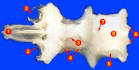
shark chondrocranium
Necturus skull
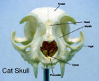
cat skull
I. SKULL
1. Work through the latter half of KZ Chapter 5 (pages 65-81) dealing with skulls.
2. Focus on the shark chondrocraniums, shark jaw set, Necturus skulls, and cat skulls. For each of these animals be able to recognize structures from the list below which are labeled in the figures and underlined in the text.
Structures to identify:
Shark:
chondrocranium
rostrum
rostral carina
precerebral cavity
rostral fenestrae
nasal capsules
nares
orbits
supraorbital crest
otic capsules
occipitum
occipital condyle
foramen magnum
splanchnocranium
mandibular arch
upper jaw
lower jaw
Meckel's cartilages
labial cartilage
branchial gill arches
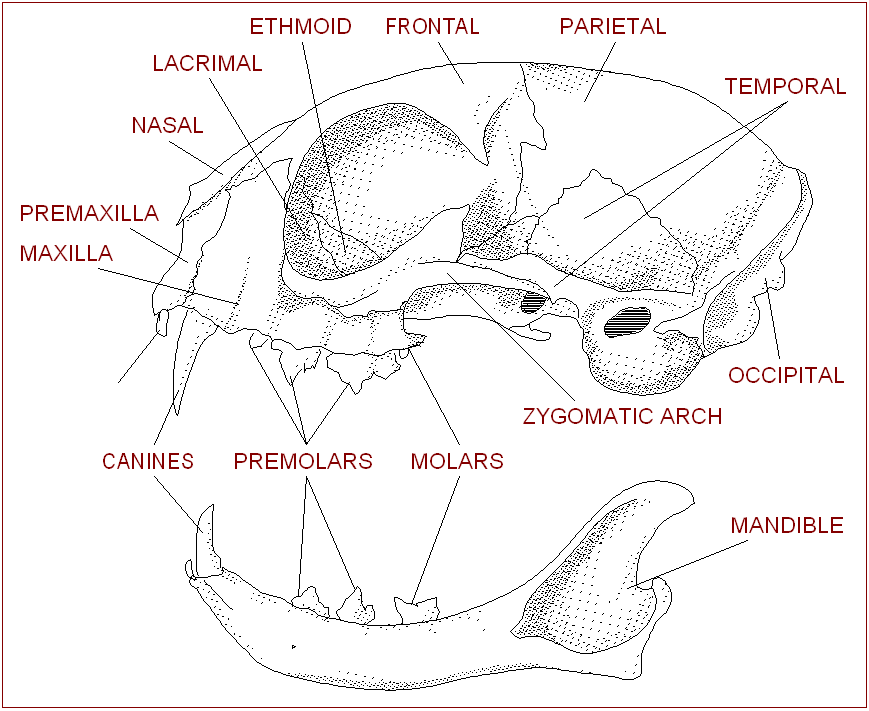
Cat:
general features
orbits
external nares
cranium
cerebral fossa
cerebellar fossa
tentorium
temporal fossae
foramen magnum
sella turcica
external auditory meatuses
tympanic bullae
bones and bony structures
nasals
premaxillae
maxillae
frontals
frontal sinus
parietals
interparietals
occipital
occipital condyles
jugal (zygomatic)
lacrimals
temporals
tympanic bullae
turbinates
palatines
vomer
ethmoid - cribriform plate
sphenoid set
sphenoidal sinus
mandibular set
(lower jaw -
several fused bones)
hyoid apparatus
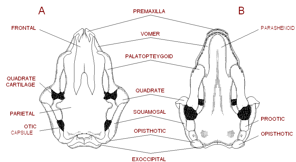
Necturus:
dermatocranium
premaxillae
frontals
parietals
quadrates
vomer
pterygoids
parasphenoid
dentary
chondrocranium
ethmiod plate
exoccipitals
foramen magnum
otic capsules
splanchnocranium
(hyoid elements are not
present on our specimens)
3) For the other vertebrates in the lab manual, be able to distinguish chondocranium, dermatocranium, and splanchnocranium contributions to the skull.
II. DENTITION
1) Work though the dentition section of KZ Chapter 5 (pages 81-83).
2) Work with the sample skulls to find examples of each of the following dentition types:
adont vs. diphyodont vs. polyphyodont
acrodont vs. pleurodont vs. thecodont
homodont vs. heterodont
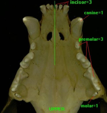
3) For each of the heterodont mammalian skulls determine the dental formula: I/I,C/C,P/P,M/M. The examples to the left show the upper and lower jaws of a lion, with the dental formula 3/3,1/1,3/3,1/0.
III. BONE AND TOOTH STRUCTURE AND DEVELOPMENT
1) Work through the slides of hyaline cartilage, elastic cartilage, fibrocartilage, and compact bone until you can recognize these tissues.
2) Work through the demonstration slides of endochondral and intramembranous bone development.
3) Identify the labeled structures on the long bone samples.
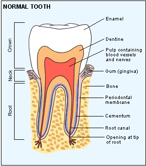
4) Examine the demonstration slide of mammalian tooth structure, using the card and the diagram link to the left as guides.
IV. TAXONOMIC KEYS
1) Classify at least two of the sample mammalian skulls to the level of Genus using the Roest guide.
2) Classify the same two sample mammalian skulls to the level of Genus using the Jones and Manning guide.
3) How well do your two sets of classifications agree?